
Pull to refresh
Logo
In-cell imaging reveals bacterial protein transport-and-folding complex

In-cell imaging reveals bacterial protein transport-and-folding complex

New Capabilities

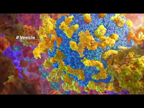
Cryo-ET maps of Mycoplasma pneumoniae show the Sec channel hitched to a dome-shaped folding chamber

Yesterday: Cell publishes the peer-reviewed study

Overview

Updated 2 hours ago

For forty years, the bacterial protein transport channel — the Sec translocon — was studied as an isolated molecule, purified out of cells and pinned in crystals. A new study watched the whole machine work inside a living bacterium and found it hitched to a dome-shaped folding chamber nobody had seen before.

The images, published September 25 in Cell, are the first sub-nanometer maps of the complete Sec machinery in its native membrane, taken in the near-minimal bacterium Mycoplasma pneumoniae. They show the ribosome passing a newborn protein straight to the channel, and three previously unknown proteins folding it into shape on the other side.

Why it matters

Seeing the full bacterial protein transport machine at work in a cell could sharpen antibiotic targets and improve engineered protein secretion.

Questions about this story

Free account needed to ask — your question is kept and asked for you right after sign-up. Answers are public.

No questions yet — be the first to ask.

Key Indicators

3
New dome proteins found
Three uncharacterized Mdps form the extracellular folding chamber.
1.8%
Ribosome-dome complexes in untreated cells
Low baseline coupling between translation and translocation.
8-16%
Ribosome-dome complexes after translation halt
Enrichment when protein synthesis is stopped with antibiotics.
Sub-nm
Resolution of in-cell maps
First sub-nanometer views of the complete Sec machinery in cells.

Voices

Curated perspectives — historical figures and your fellow readers.

Ever wondered what historical figures would say about today's headlines?

Sign up to generate historical perspectives on this story.

People Involved

Organizations Involved

Timeline

April 2025 September 2026

2 events Latest: Yesterday
  1. Cell publishes the peer-reviewed study

    Latest Publication

    The paper appears online with sub-nanometer in-cell maps of the complete Sec translocation-folding complex.

  2. Preprint posted on bioRxiv

    Research

    The team reports the discovery of a translocation-folding complex in Mycoplasma pneumoniae before peer review.

Scenarios

1

Dome-folding complex found in other bacteria

Likely Resolves by Q2 2028

Discussed by: The study authors, who note whether similar mechanisms exist in other organisms remains to be explored

Cryo-ET screens of other bacteria — model organisms like E. coli or pathogens — reveal homologous dome complexes coupled to the Sec translocon. A structure-first study using the same pipeline on a second species would settle it.

2

Dome components engineered to boost protein secretion

Possible Resolves by End of 2028

Discussed by: Structural biology and synthetic biology commentators covering the paper

Researchers graft the dome folding chamber onto engineered secretion systems to improve yields of recombinant proteins in bacterial production. The three Mdps and their foldase activity become a biotech tool.

3

SecA co-translational handoff confirmed in E. coli

Possible Resolves by Q2 2028

Discussed by: The paper's authors, citing growing E. coli evidence for SecA-mediated co-translational translocation

Follow-up experiments confirm the model in E. coli, showing ribosomes hand nascent peptides to SecA physically across a wider range of bacteria. The current maps are the first direct in-cell evidence for this handoff.

Historical Context

2 moments from history that rhyme with this story — and how they unfolded.

1999–2009

Ribosome crystal structures (2000s)

X-ray crystallography produced atomic models of the ribosome, the cell's protein-making machine, after decades of effort. The 2009 Nobel Prize in Chemistry went to Venki Ramakrishnan, Thomas Steitz, and Ada Yonath for the work.

Then

Structural biology gained its most detailed view of a core cellular machine.

Now

Ribosome structures became a foundation for antibiotics research and a template for studying other complexes.

Why this matters now

The same ambition — resolve a cellular machine at atomic clarity — is now achieved inside the cell with cryo-ET, without purifying the complex first.

early 2010s

Nuclear pore complex in-cell cryo-ET (2010s)

Martin Beck's group at EMBL used cryo-electron tomography to build integrative models of the nuclear pore complex, the gatekeeper between nucleus and cytoplasm, from images of intact cells rather than purified samples.

Then

It became a landmark proof that large complexes could be resolved in their native context.

Now

It helped establish cryo-ET as in-cell structural biology and trained the methods used today.

Why this matters now

Mahamid's group, working in the same EMBL setting, extends that approach to a completely new machine.

Sources

(6)