
Pull to refresh
Logo
In-cell imaging reveals how bacteria coordinate protein production and transport

In-cell imaging reveals how bacteria coordinate protein production and transport

New Capabilities

Cryo-electron tomography maps 140 translation complexes inside Mycoplasma pneumoniae, linking transcription to membrane transport

Yesterday: Cell publishes two companion papers

Overview

Updated 2 hours ago

For decades, structural biologists had to purify molecules before they could image them. The ribosome's structure was solved from isolated particles, frozen in test tubes. Two papers in Cell now show the same machinery at work inside intact bacterial cells, exposing coordination that was only theorized before.

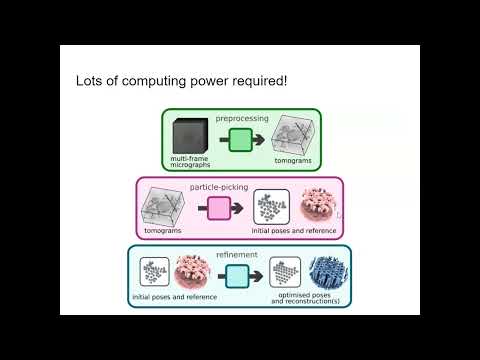
The team, led by Julia Mahamid at EMBL Heidelberg, used cryo-electron tomography on Mycoplasma pneumoniae, a bacterium with one of the smallest genomes known. They resolved 140 structures spanning every phase of translation and found a supercomplex linking transcription, translation, and membrane transport. A companion paper maps the protein channel that pushes new proteins across the membrane, plus a dome of three newly identified proteins that fold them as they exit.

Why it matters

Scientists can now watch protein production inside intact cells, revealing coordination mechanisms that appear conserved from bacteria to humans.

Questions about this story

Free account needed to ask — your question is kept and asked for you right after sign-up. Answers are public.

No questions yet — be the first to ask.

Key Indicators

140
Translation complex maps resolved
Maps cover initiation, elongation, termination, and recycling phases in native cells.
3
Previously unknown proteins in the membrane translocation complex
Called Mdps, they form an extracellular dome that folds proteins as they leave the cell.
Sub-nanometer
Resolution of complete bacterial Sec-translocation machinery
First sub-nanometer maps of the translocon, including the attached folding dome.

Voices

Curated perspectives — historical figures and your fellow readers.

Ever wondered what historical figures would say about today's headlines?

Sign up to generate historical perspectives on this story.

People Involved

Organizations Involved

Timeline

October 2025 September 2026

2 events Latest: Yesterday
  1. Cell publishes two companion papers

    Latest Publication

    The visual proteomics study and the Sec-translocon complex paper appear online in Cell, revealing the transcription-translation-membrane supercomplex.

  2. Preprint posted to bioRxiv

    Research

    Dobbs and colleagues post the single-cell visual proteomics study of Mycoplasma pneumoniae translation to bioRxiv.

Scenarios

1

Membrane-ribosome mechanism confirmed in mammalian cells

Possible Resolves by End of 2028

Discussed by: The Cell paper's discussion; Phys.org notes the mechanism mirrors behavior seen in mammalian cells decades ago.

The study found large ribosomal subunits sitting on the cell membrane even when idle, detaching only when a new round of translation can begin. That behavior was observed in mammalian cells decades ago, but never structurally explained. A follow-up testing this directly in mammalian cells would confirm the mechanism is conserved across kingdoms.

2

Antibiotic-perturbation maps yield new drug targets

Possible Resolves by End of 2027

Discussed by: The study itself, which imaged antibiotic-treated cells; Phys.org notes that understanding these processes informs drug development.

The team imaged cells treated with antibiotics that stall translation. Those maps show where drugs interrupt the coupling of transcription and translation. A follow-up identifying a compound that disrupts the newly discovered supercomplex would open a fresh antibacterial target.

3

In-cell gene-expression mapping expands beyond minimal bacteria

Likely Resolves by Q2 2028

Discussed by: Grant Jensen's comments on applying the methods to complex organisms; Julia Mahamid's remarks on studying molecular communities.

The cryo-ET workflow developed here, single-cell visual proteomics, could be applied to other bacteria or to human cells. A published application to a non-minimal organism would validate the approach beyond Mycoplasma.

Historical Context

2 moments from history that rhyme with this story — and how they unfolded.

2010–2016

JCVI minimal genomes (2010, 2016)

The J. Craig Venter Institute built synthetic genomes and transplanted them into cells, creating first JCVI-syn1.0 (2010) and later JCVI-syn3.0 (2016) and syn3A. Syn3A carries 493 genes on a single 543 kilo-base-pair chromosome, among the smallest genomes capable of supporting growth.

Then

A minimal-cell platform for defining the gene set required for life.

Now

Genome-reduced Mycoplasma species became the model of choice for studying basic cellular processes.

Why this matters now

M. pneumoniae's small genome and simple architecture make it ideal for the in-cell imaging shown in the Cell papers.

2013–2017

Cryo-EM resolution revolution (2013–2017)

Direct electron detectors and new computational methods pushed cryo-electron microscopy to near-atomic resolution, letting scientists solve structures of proteins too large or fragile for X-ray crystallography. The 2017 Nobel Prize in Chemistry went to Jacques Dubochet, Joachim Frank, and Richard Henderson for developing cryo-EM.

Then

A flood of high-resolution structures of ribosomes, ion channels, and viral capsids.

Now

Structural biology shifted from protein crystals to single particles, but still relied on purified molecules.

Why this matters now

The Cell studies extend that revolution one step further, imaging the same machines inside intact cells without purification.

Sources

(6)