

Pull to refresh
Logo
Studies find brain cell axons are strings of pearls, not smooth tubes

Studies find brain cell axons are strings of pearls, not smooth tubes

New Capabilities

Two studies, in mouse and human brain tissue, show nerve cell extensions pearled along their length with shapes that change as neurons fire.

Today: ScienceDaily synthesizes pearled axon findings

Overview

Updated 1 hour ago

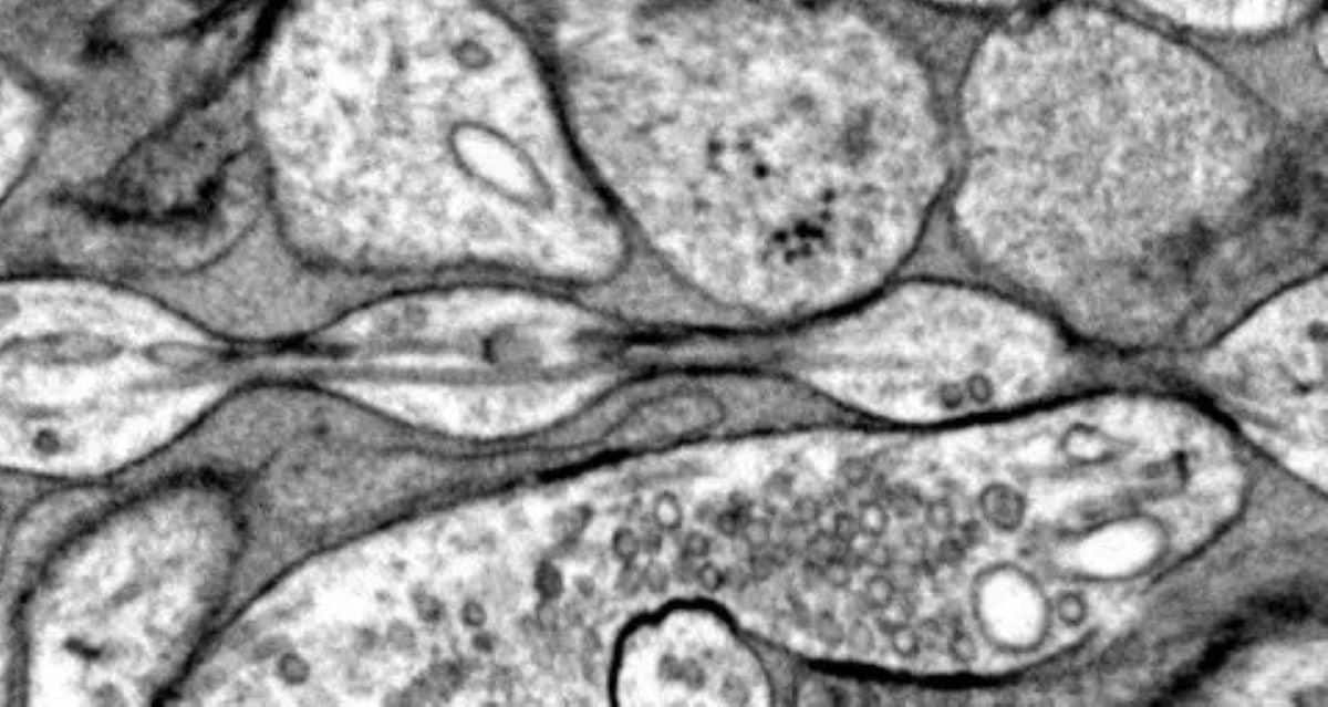
Textbook diagrams have drawn brain cells with smooth, tube-like extensions for more than a century. Researchers at Johns Hopkins University found that many of those extensions actually resemble strings of tiny pearls, with repeating bulges that change size as neurons fire.

The shape appears to influence how quickly electrical signals travel, which could mean the brain has a mechanical way to tune its own communication speed. It also blurs the line between normal variation and the beading long associated with brain damage.

Why it matters

If axon shape tunes signal speed, the brain has a mechanical dial for communication that textbooks missed for a century.

Questions about this story

Free account needed to ask — your question is kept and asked for you right after sign-up. Answers are public.

No questions yet — be the first to ask.

Key Indicators

250 nm
Diameter of pearl-like swellings on axons
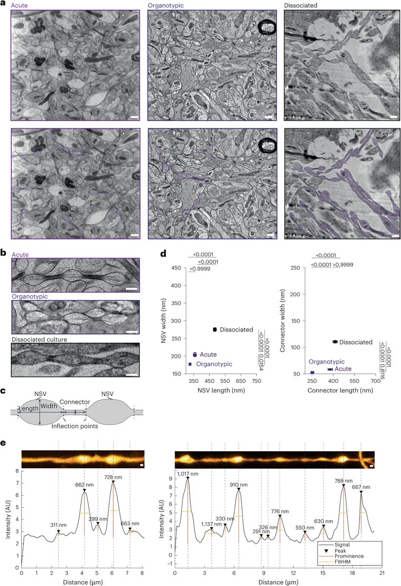
Repeating bulges about 250 nanometers across, connected by segments about 70 nanometers wide.
17%
Width increase of pearls after high-frequency stimulation
Pearls widened 17% and lengthened 8% after stimulation, with enlargement lasting at least 30 minutes.
100+ years
How long textbooks have shown axons as smooth tubes
Standard neuron drawings trace back to Santiago Ramón y Cajal's late-1800s illustrations.

Voices

Curated perspectives — historical figures and your fellow readers.

Ever wondered what historical figures would say about today's headlines?

Sign up to generate historical perspectives on this story.

People Involved

Organizations Involved

Timeline

December 2024 September 2026

4 events Latest: Today
Tap a bar to jump to that date
  1. ScienceDaily synthesizes pearled axon findings

    Today Science communication

    ScienceDaily publishes a consolidated report on both studies, highlighting activity-dependent reshaping and open questions about disease.

  2. Human tissue study appears in Neuron print issue

    Research publication

    The human confirmation study is published in the journal's February issue.

  3. Human brain tissue confirms pearled axons

    Research publication

    Eddings-led study in Neuron shows pearled axons in cortical tissue from epilepsy surgeries, extending findings beyond mice.

  4. Mouse axon pearling described in Nature Neuroscience

    Research publication

    Watanabe lab reports non-synaptic varicosities along nonmyelinated mouse axons, with mathematical models and cholesterol experiments linking membrane tension to pearling.

Scenarios

1

Pearling confirmed in myelinated axons and tied to signal-speed control

Likely Resolves by Q2 2028

Discussed by: Watanabe lab research plans; ScienceDaily reporting

The original mouse study examined only nonmyelinated axons, leaving open whether pearling occurs in myelinated fibers. A follow-up demonstrating activity-dependent pearling with direct conduction-velocity measurements would settle whether the shape genuinely regulates signal timing across the brain.

2

Abnormal pearling identified as a biomarker in neurological disease

Possible Resolves by Q2 2029

Discussed by: Watanabe lab's planned examination of tissue from people who died with neurodegenerative diseases

The team plans to compare pearling patterns in healthy tissue against tissue from patients with neurodegenerative diseases. If specific pearling abnormalities track a named condition, the structure could become a diagnostic marker and a way to separate normal variation from disease-related damage.

3

Pearling shown to be a preparation artifact with no signaling role

Unlikely Resolves by End of 2027

Discussed by: ScienceAlert called the 2024 study 'controversial'; replication question remains open

Critics could argue the pearling emerges from tissue preparation or imaging rather than existing in living brains. In-vivo imaging showing smooth axons, or experiments demonstrating no conduction change with pearling, would undercut the signaling claim. The human tissue confirmation makes this scenario less likely, but it cannot be ruled out until direct in-vivo evidence appears.

Historical Context

2 moments from history that rhyme with this story — and how they unfolded.

1888–1959

Discovery of dendritic spines (1888–1950s)

Ramón y Cajal first drew small protrusions on dendrites, but many scientists dismissed them as staining artifacts. Only in the 1950s, with electron microscopy, did researchers confirm spines are real, dynamic structures that house synapses and change with learning.

Then

Electron microscopy settled the artifact debate and confirmed spines as genuine anatomy.

Now

Dendritic spines are now central to understanding synaptic plasticity, learning, and memory.

Why this matters now

Like the spines, the pearled axon varicosities were initially described through painstaking imaging, and only independent replication will determine whether they earn a permanent place in the anatomical canon.

Late 1890s–1906

Neuron doctrine debate, Golgi vs. Cajal (1906)

Camillo Golgi and Santiago Ramón y Cajal shared the 1906 Nobel Prize yet disagreed on the most basic question of brain structure: whether neurons were physically continuous or separate cells. Cajal's drawings of discrete neurons, using Golgi's own staining method, eventually won.

Then

Cajal's neuron doctrine became the foundation of modern neuroscience.

Now

Textbook illustrations based on Cajal's drawings shaped how generations of students visualized the brain.

Why this matters now

The pearled axon finding challenges the same textbook lineage, suggesting Cajal's smooth-tube drawings, however elegant, omitted a structural feature visible only with modern imaging.

Sources

(5)