
Pull to refresh
Logo
Prefrontal cortex mapped at single-cell resolution across the full human lifespan

Prefrontal cortex mapped at single-cell resolution across the full human lifespan

New Capabilities

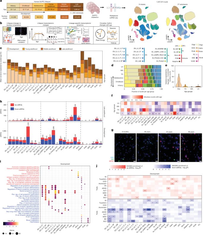
1.3 million nuclei from 284 brains reveal three transcriptomic acts: development, stability, and late-life glial reactivation

2 days ago: Atlas and nine companion studies published in Nature

Overview

Updated 2 hours ago

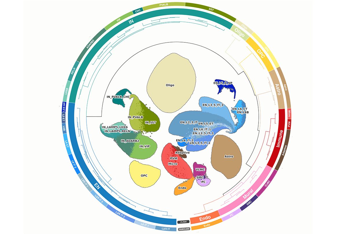
Scientists have mapped how every major cell type in the human prefrontal cortex changes across the entire lifespan — from birth to age 97. The atlas, published in Nature on September 23, 2026, profiles 1.3 million nuclei from 284 neurotypical donors and reveals that the brain's molecular activity proceeds in three distinct acts: intense remodeling through adolescence, relative stability through midlife, and a late-life surge of change concentrated in glial cells.

The finding reframes how researchers think about brain aging. Neurons, long assumed to be the most fragile cells, are actually transcriptionally stable well into old age. It's the support cells — microglia, astrocytes, and oligodendrocytes — that show dramatic molecular reactivation after age 60, converging on immune activation, stress responses, and disrupted circadian rhythms. That shift, the authors argue, could be the earliest molecular signature of age-related cognitive decline and disease vulnerability.

Why it matters

This atlas gives researchers a normative baseline for brain aging — without it, they can't distinguish healthy trajectories from disease onset in Alzheimer's, Parkinson's, or schizophrenia.

Questions about this story

Free account needed to ask — your question is kept and asked for you right after sign-up. Answers are public.

No questions yet — be the first to ask.

Key Indicators

1.3M
Nuclei profiled in the DLPFC atlas
Single-nucleus transcriptomes from 284 postmortem samples spanning ages 0-97.
284
Neurotypical donors
Postmortem samples ranging from infancy to 97 years old.
24
Age of compositional inflection point (years)
After age 24, most neuronal and glial subclass composition stabilizes sharply.
6.3M
Total cells across all nine Nature studies
Cells from 1,494 deceased donors, ages infancy to 108, various ancestries.

Voices

Curated perspectives — historical figures and your fellow readers.

Ever wondered what historical figures would say about today's headlines?

Sign up to generate historical perspectives on this story.

People Involved

Organizations Involved

Timeline

November 2024 September 2026

5 events Latest: 2 days ago
Tap a bar to jump to that date
  1. Atlas and nine companion studies published in Nature

    Latest Publication

    Nature published the lifespan atlas plus eight companion studies analyzing 6.3 million cells from 1,494 donors, spanning development, aging, and disease.

  2. Researchers find age 24 is a stability inflection point

    Finding

    The atlas showed most cell-type composition stabilizes sharply after age 24, except SST-positive interneurons and oligodendrocyte precursor cells.

  3. Circadian disruption identified in older adult brains

    Finding

    Daily clock genes showed coordinated patterns in younger neurons but weakened, desynchronized patterns in older adults; glia gained stress-associated rhythms.

  4. Glial reactivation mapped to late-life immune and stress programs

    Finding

    Microglia, astrocytes, and oligodendrocytes converged on immune activation and unfolded protein response genes after age 60, while neurons stayed transcriptionally stable.

  5. Lifespan atlas preprint posted on medRxiv

    Publication

    The full DLPFC lifespan atlas, 1.3 million nuclei from 284 donors ages 0-97, appeared as a preprint.

Scenarios

1

Glial aging programs become validated drug targets for Alzheimer's and Parkinson's

Possible Resolves by Sep 23, 2027

Discussed by: Roussos and other PsychAD researchers quoted in Reuters coverage

The atlas identifies late-life glial immune and stress programs as the earliest molecular shifts in the prefrontal cortex. Researchers at Mount Sinai and partners use these programs to screen for compounds that dampen microglial activation or restore oligodendrocyte function. A scaled validation study in animal models or organoids follows. If successful, this produces a named therapeutic candidate entering clinical trials.

2

Atlas becomes the standard normative reference for prefrontal cortex research

Likely Resolves by Sep 23, 2027

Discussed by: Nature news editors; companion studies by Yang et al., Lee et al., Venkatesh et al.

The 284-donor lifespan resource is adopted as the default healthy baseline for interpreting disease-affected brain transcriptomes. Subsequent single-cell studies of Alzheimer's, schizophrenia, and aging begin citing it as the normative control set, mirroring how the Allen Brain Atlas became a standard reference. New tools and reanalysis packages built on the published data appear within the year.

3

Circadian disruption findings spawn interventional trials in older adults

Possible Resolves by Sep 23, 2027

Discussed by: The Transmitter coverage highlighting circadian reorganization as a novel finding

The observed loss of neuronal clock synchronization in older adults becomes a testable hypothesis. A clinical group designs a pilot trial of a circadian intervention — timed light exposure, melatonin, or clock-gene modulators — measuring molecular markers in accessible tissues or imaging readouts. The atlas provides the normative molecular signature to track whether intervention restores synchronization.

Historical Context

3 moments from history that rhyme with this story — and how they unfolded.

2006

Allen Brain Atlas (2006)

The Allen Institute released a genome-wide expression map of the adult mouse brain, profiling roughly 20,000 genes across the entire brain at cellular resolution. It became the first comprehensive, open-access reference for where genes are active in a mammalian brain.

Then

Neuroscientists gained a standard spatial reference for gene expression, accelerating studies of brain function and disease.

Now

The Allen Brain Atlas became a foundational tool cited in tens of thousands of papers and spawned the Human Brain Atlas and other follow-on references.

Why this matters now

The DLPFC lifespan atlas follows the same pattern — building an open reference dataset that the field adopts as a normative baseline. Unlike the Allen Atlas's spatial focus, this one adds the dimension of time: how every cell type changes from birth to old age.

2016-Present

Human Cell Atlas launches (2016)

An international consortium launched the Human Cell Atlas with the goal of mapping every cell type in the human body using single-cell genomics. It organized a massive, open-data effort across hundreds of labs to characterize the molecular states of cells across tissues and developmental stages.

Then

The effort produced standardized protocols, data-sharing frameworks, and cell-type taxonomies that made large-scale atlases technically feasible.

Now

It established the norms of open reference atlases and data reuse that the PsychAD lifespan atlas now extends to the aging brain.

Why this matters now

The long lifespan (0-97 years) and the focus on postmortem normative tissue distinguish this from the Human Cell Atlas's tissue-type focus. The DLPFC atlas adds a temporal dimension that the HCA's early efforts lacked.

2019-2023

Early single-cell Alzheimer's transcriptomic studies (2019-2023)

Multiple groups published single-nucleus transcriptomic studies of Alzheimer's-affected brains, identifying disease-associated microglia and astrocyte states. Most used small donor cohorts or restricted age ranges, making it hard to separate disease effects from normal aging.

Then

Disease-associated cell states were identified, but ambiguous whether they were causes, consequences, or aging artifacts.

Now

These studies created the questions the lifespan atlas now answers: which molecular changes are normative aging, and which are pathological.

Why this matters now

The new atlas's 284 neurotypical donors provide the missing normative baseline. Researchers can now ask whether an Alzheimer's-associated microglial state is a disease-specific signature or an accelerated version of normal aging.

Sources

(8)