

Pull to refresh
Logo
First spatial map of mouse brain lipids reveals hundreds of biochemical zones

First spatial map of mouse brain lipids reveals hundreds of biochemical zones

New Capabilities

EPFL team maps 172 lipids across the mouse brain, uncovering zones tied to cell types, wiring, and pregnancy

2 days ago: Nature publishes the brain lipid atlas

Overview

Updated 1 hour ago

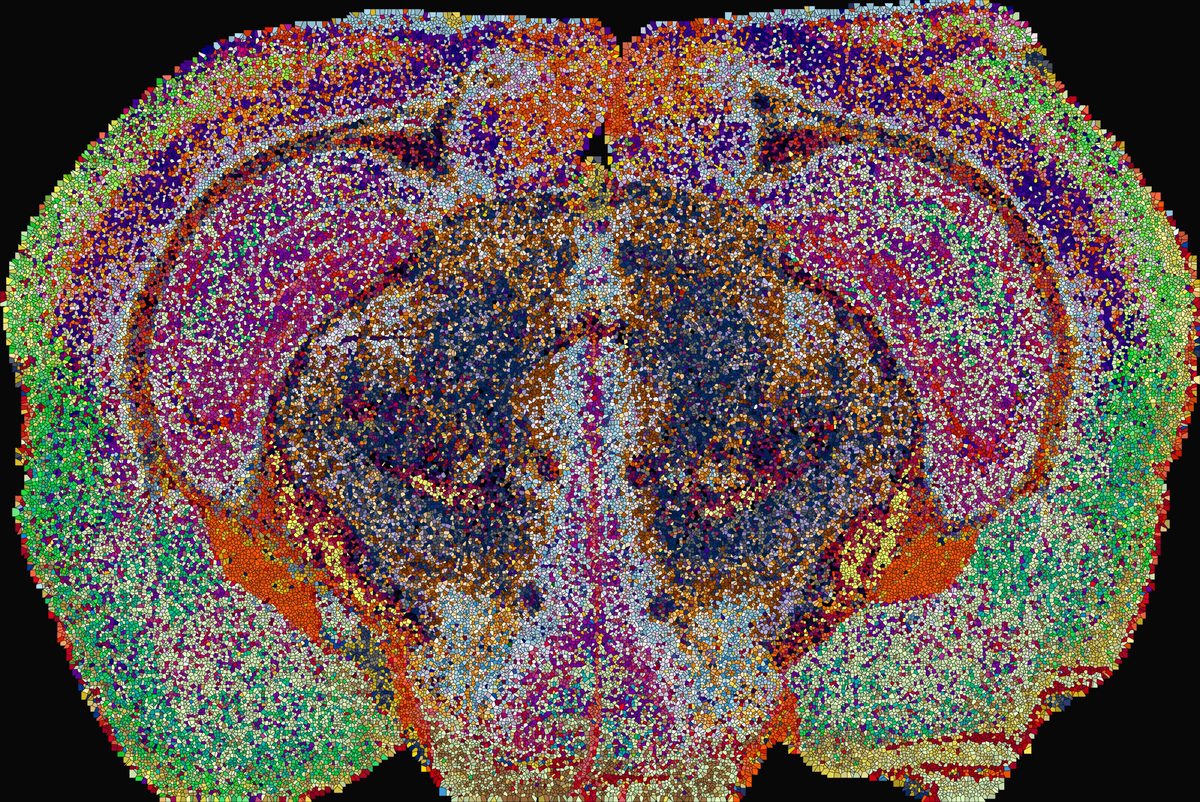
Researchers at EPFL mapped the lipid chemistry of the entire mouse brain, dividing it into 539 biochemical territories they call lipizones. Published in Nature on September 23, the atlas links lipid composition to cell types, distant axon terminals, and brain connectivity.

The map also captures pregnancy's effect on brain lipids. Changes were larger than typical sex differences, with the cortex reshaped at layer 4 and a myelin lipid rising across 100 of 215 lipid supertypes. It is the first spatial reference of its kind for studying how lipids shape brain function and disease.

Why it matters

Brain lipid chemistry can now be tied to specific territories and wiring, opening a new diagnostic and therapeutic lens on myelin disease and Alzheimer's.

Questions about this story

Free account needed to ask — your question is kept and asked for you right after sign-up. Answers are public.

No questions yet — be the first to ask.

Key Indicators

539
Lipizones (biochemical territories) identified
Each lipizone has a signature mix of membrane lipids, mapped at cell-scale resolution.
172
Lipid species mapped across the brain
Mapped from 109 coronal sections from 11 mice using MALDI mass spectrometry imaging.
76%
Lipizones matching known cell-type territories
304 of 399 lipizones overlapped cell-type territories defined by other methods.
7.2M
Pixels in the imaging dataset
26,874 mass peaks were detected across the full brain volume.

Voices

Curated perspectives — historical figures and your fellow readers.

Ever wondered what historical figures would say about today's headlines?

Sign up to generate historical perspectives on this story.

People Involved

Organizations Involved

Timeline

October 2025 September 2026

2 events Latest: 2 days ago
  1. Nature publishes the brain lipid atlas

    Latest Publication

    Peer-reviewed paper describes 539 lipizones, connectivity-linked lipid signatures, oligodendrocyte heterogeneity, ventricular zonation, and pregnancy-driven remodeling.

  2. Preprint of the lipid atlas posted on bioRxiv

    Publication

    The team shared their full mouse brain lipid mapping work as a preprint ahead of peer review.

Scenarios

1

Lipid atlas becomes neuroscience standard reference

Likely Resolves by End of 2028

Discussed by: The authors, who describe the work in Nature as 'a foundational resource'

Research groups adopt lipizones as a standard unit of brain organization, layering lipids onto existing atlases of gene expression and connectivity. The MALDI-MSI approach spreads to other labs, species, and disease models, much as the Allen Brain Atlas became a standard template.

2

Human studies confirm pregnancy brain lipid changes

Possible Resolves by End of 2029

Discussed by: The authors, who note pregnancy is associated with lower Alzheimer's risk in women

A human post-mortem or imaging study finds comparable lipid shifts tied to pregnancy, connecting the mouse findings to women's brain health. Such a result would strengthen the hypothesis linking parity, lipid remodeling, and reduced dementia risk, opening a new avenue for Alzheimer's prevention research.

3

Atlas accelerates lipid-targeting drug development

Uncertain Resolves by End of 2029

Discussed by: The paper identifies specific lipids and genes tied to myelin biology and cortical remodeling

Pharmaceutical or biotech companies shortlist lipid pathways, especially sphingolipids and galactosylceramide, as targets for myelin repair or Alzheimer's disease. A named candidate enters clinical testing, or a licensing partnership is announced using atlas data to justify the approach.

Historical Context

3 moments from history that rhyme with this story — and how they unfolded.

2004–2013

Allen Brain Atlas (2004–2013)

The Allen Institute mapped gene expression across the mouse brain, using in situ hybridization to catalog thousands of genes at cellular resolution. The effort produced a publicly accessible, standardized reference of brain gene activity.

Then

The atlas became a standard resource, cited in thousands of papers within a decade.

Now

It established the template for systematic molecular atlasing of the brain, later extended to human tissue.

Why this matters now

The lipid atlas follows the same model, adding a biochemistry layer that the gene-based Allen atlas could not capture.

2009–2019

Human Connectome Project (2009–2019)

The project mapped white matter tracts and functional connections in the human brain using diffusion MRI and resting-state imaging. It produced wiring diagrams of the healthy adult brain.

Then

Connectivity maps became standard tools for studying brain networks and disorders.

Now

The wiring framework now underpins thousands of studies linking structure to function and disease.

Why this matters now

The lipid atlas shows connected brain regions share lipid signatures, suggesting lipids add a biochemical dimension to connectivity mapping.

2016–2020

Rise of spatial transcriptomics (2016–2020)

Methods like MERFISH and seqFISH began mapping RNA molecules directly in intact tissue, revealing cell types and states in their native spatial context. The Human Cell Atlas and Tabula Muris projects applied these tools across organs.

Then

Spatial transcriptomics reshaped cell classification by adding location to gene expression data.

Now

It created the precedent that spatial molecular maps are essential reference tools for biology.

Why this matters now

The lipid atlas extends this spatial mapping approach from gene expression to lipid chemistry, the first such reference for any organ.

Sources

(7)