
Pull to refresh
Logo
MIT researchers build handheld device that collects living cancer cells for testing

MIT researchers build handheld device that collects living cancer cells for testing

New Capabilities

A fluid-flow tool harvests live cells from tissue without the chemical preservation that kills them, enabling organoid growth and personalized drug testing

Yesterday: Study describing cell-collection device published in Device

Overview

Updated Yesterday

Standard cancer pathology has a built-in contradiction: tissue is bathed in chemical preservatives before analysis, and those chemicals kill the cells inside it. That leaves pathologists with a good look at dead cells and no way to test how a patient's living cancer responds to drugs.

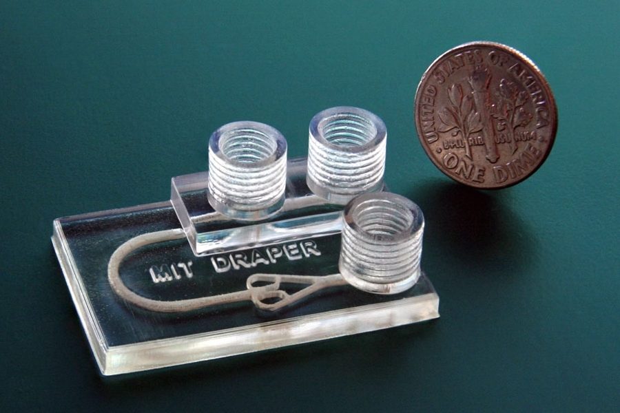
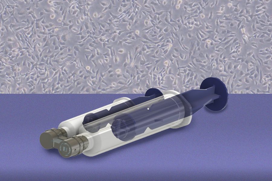
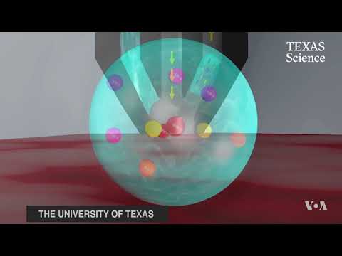
MIT researchers, with collaborators at Johns Hopkins and Princeton, built a handheld device that breaks that tradeoff. A 3D-printed microfluidic tool holds a tiny channel against fresh tissue and pushes fluid across the surface, using shear force to detach living cells from a pinpoint location without cutting or chemically treating the sample.

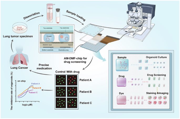
The collected cells survive and can be grown into organoids, miniature lab models of a patient's disease, then screened against therapies. The team plans first to use the device on tissue already removed during surgery, with the longer-term goal of sampling cells inside the body for earlier detection.

Why it matters

If it reaches clinics, doctors could test how a patient's living cancer cells respond to drugs, and detect cancer earlier without destroying tissue.

Questions about this story

Free account needed to ask — your question is kept and asked for you right after sign-up. Answers are public.

No questions yet — be the first to ask.

Key Indicators

1–5 pascals
Shear stress range to detach different cell types
Loosely adherent prostate cancer cells detach at 1 pascal; bone cancer cells need up to 5 pascals.
0
Chemicals or enzymes added during collection
The flow-based method needs no chemical or enzymatic treatment, preserving cell viability.
Human
Tissue type the device was tested on
Fresh human fallopian tube samples shipped from Johns Hopkins yielded cells that grew into organoids.

Voices

Curated perspectives — historical figures and your fellow readers.

Ever wondered what historical figures would say about today's headlines?

Sign up to generate historical perspectives on this story.

People Involved

Organizations Involved

Timeline

1 event Latest: Yesterday
  1. Study describing cell-collection device published in Device

    Latest Research Publication

    MIT and collaborators published a handheld microfluidic device that collects living cells from fresh tissue via fluid shear, enabling organoid growth and personalized drug testing.

Scenarios

1

Handheld cell-collection device enters clinical trials

Possible Resolves by End of 2028

Discussed by: The MIT research team, which said ex-vivo use offers an easier path to regulatory approval

The team's stated near-term plan is to validate the device on tissue already removed from the body, starting with fallopian tube samples for ovarian cancer detection. If a clinical partner and funding emerge, the device could be tested against standard pathology on surgical specimens and move toward regulatory clearance.

2

In-vivo sampling demonstrated inside patients

Unlikely Resolves by End of 2030

Discussed by: The MIT team, which called in-body swabbing the longer-term goal for earlier cancer detection

The long-term aspiration is a version of the device that samples tissue inside the body, likely through an endoscopic or catheter-based design. This requires proving the vacuum-sealed microfluidic sampling works safely on living tissue and clearing substantial regulatory hurdles.

3

Technology remains a research tool

Possible Resolves by End of 2031

Discussed by: Implicit in the study, which emphasizes research uses such as studying carcinogenesis

Diagnostic devices face long regulatory timelines and high validation costs. If no clinical partner or licensee emerges, the device could stay in academic labs, used to grow organoids and study disease biology but not reaching patient care.

Historical Context

3 moments from history that rhyme with this story — and how they unfolded.

2009

Organoid revolution (2009)

Hans Clevers and colleagues at the Hubrecht Institute showed that a single intestinal stem cell could grow into a self-organizing organoid, launching a field of personalized disease modeling built on living cells.

Then

Organoids became a standard tool for drug testing and disease research.

Now

They depend on living cells, which standard clinical pathology destroys with chemical preservatives.

Why this matters now

The MIT device's central value is that it supplies the living cells organoids require, connecting surgical specimens to personalized medicine.

September 2017

MasSpec Pen (2017)

Researchers at the University of Texas at Austin, including Livia Eberlin, published work on a handheld MasSpec Pen that touched tissue with a water droplet and used mass spectrometry to distinguish cancer from healthy tissue in seconds. It was tested on tissue samples, including from patients with ovarian cancer.

Then

Demonstrated rapid, nondestructive cancer identification, with the team pursuing clinical translation.

Now

Showed a small, hand-guided device could aid cancer detection without a scalpel, spurring similar diagnostic tools.

Why this matters now

The MasSpec Pen detects chemical signatures of cancer. The new MIT device instead collects living, culturable cells from tissue, pursuing earlier and less destructive cancer assessment by a different mechanism.

2019

Circulating tumor cell capture (2019)

MIT researchers published in the Proceedings of the National Academy of Sciences a microfluidic system that collected circulating tumor cells (CTCs) from mice over days and even weeks, allowing analysis as disease progressed.

Then

Enabled longitudinal study of metastasis from blood, with ambitions in liquid biopsy.

Now

Extended the idea of live cancer cell capture, though from blood rather than solid tissue.

Why this matters now

The new device applies the same live-cell capture concept to solid tissue while leaving the source sample intact for pathology.

Sources

(6)